Smart Projects : The Association launches a new medical program!
We have developed the "Smart Projects" program, which is part of our goal to constantly improve the child's care pathway. With this program, we are seeking to make progress in diagnosis using the latest Medtech innovations. The ultimate goal is to improve the management of complex heart diseases.
A team of doctors, including cardiopediatricians and cardiac surgeons, meets every week to review the medical files we receive.
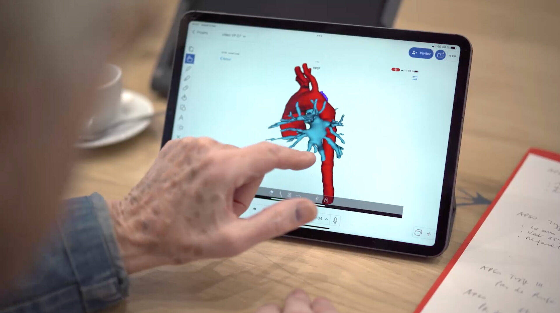
During the Covid-19 pandemic, our teams found it difficult to obtain complete medical files containing all the examinations required for decision-making. That's why Pr Francine Leca, founder of the Association, wanted to improve this part of the child's journey by using technology to serve the medical profession. The result is the first project in the Smart Projects program, based on 3D modeling, in collaboration with the start-up Visible Patient.
3 questions about the 3D modeling project:
1. What's involved in working with Visible Patient?
Visible Patient is the first online laboratory for 3D modeling of medical images. Starting with a CT scan, they transform the image into a perfect 3D visualization of the patient's internal anatomy.
2. How does Mécénat Chirurgie Cardiaque use this technology?
With the help of a CT scan of the child taken in his own country, and its 3D modeling, doctors in France are able to make an accurate diagnosis of the heart disease.
3. What are the advantages of 3D modeling?
- Confirm diagnosis and precise cardiac anatomy
- Precisely plan the surgical procedure to be performed whenever possible
- Teaching, by exchanging case studies with foreign doctors.
The case of APSO
Every year, the association cares for around 75 children (25%) suffering from complex cardiopathies. Among these rare and serious cardiopathies is APSO (Open Septum Pulmonary Atresia). To successfully operate on this malformation, it is essential to first assess the patient's pulmonary tree. Until now, this could only be done by angiography performed during cardiac catheterization, an examination to which the children we treat do not have access in their own countries. Today, with the support of a good CT scan, 3D reconstruction provides us with essential information on the morphology of pulmonary vascularization.
To begin with, we naturally chose to focus our 3D modeling on patients with APSO, and this has borne fruit: we have accepted files from patients whose pulmonary branches were sufficiently developed to allow them to be managed, and we have avoided unnecessarily bringing in those whose cardiac anatomy did not allow surgical intervention.
What's next?
As 3D modeling is based on a scanner, we are currently working with 13 countries equipped with the right equipment. In the future, we hope to enable as many children as possible to benefit from this technology.